3D Intra-Cardiac Echography (3D-ICE)
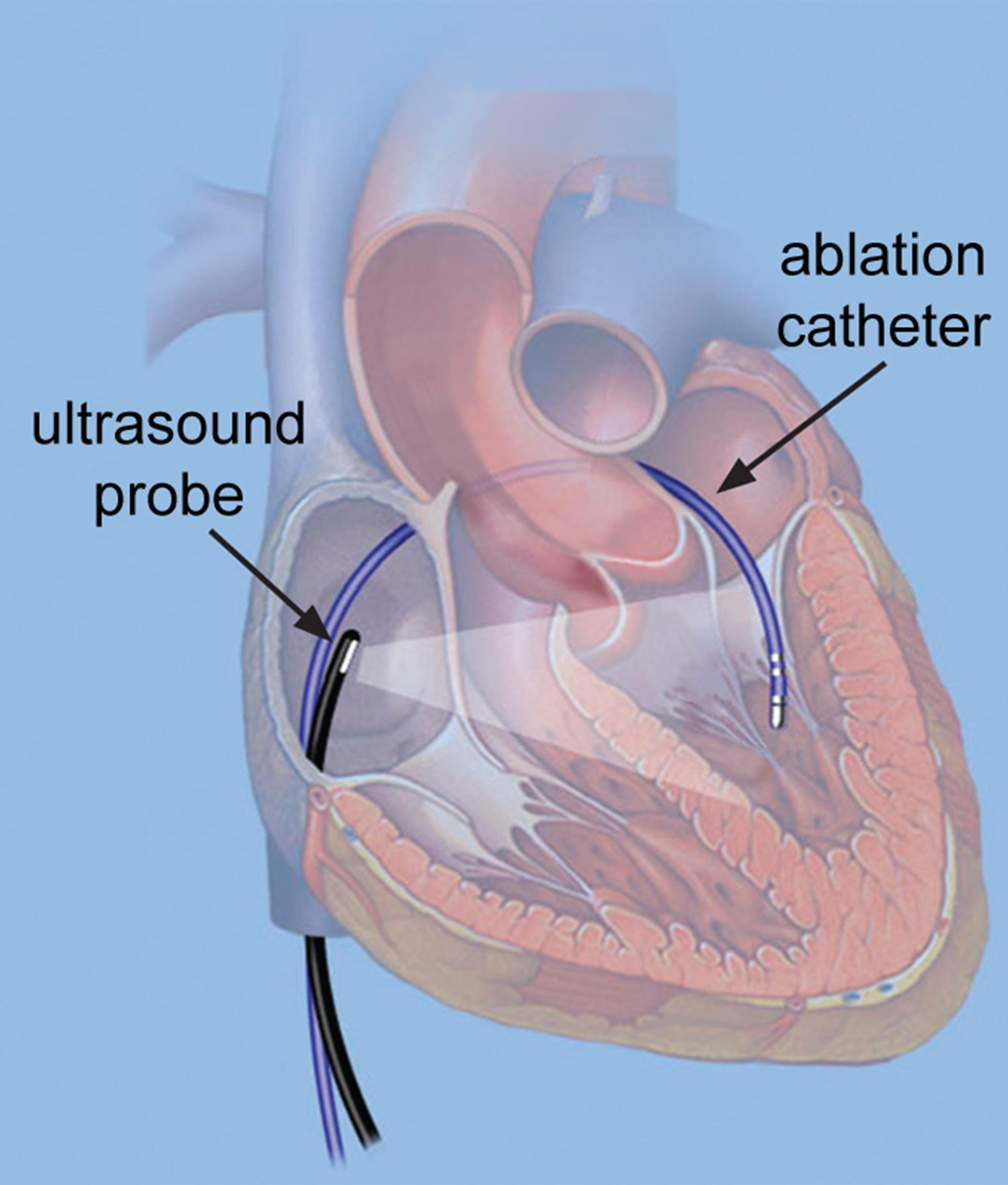
A variety of cardiovascular conditions can be treated using minimally-invasive interventions. These interventions are much less stressful to the patient than conventional surgery, shortening recovery time and allowing treatment to take place even at advanced age. Examples are rhythm-disorder treatments or catheter-based valve replacement. Accurate visualization is crucial for the success of such interventions, but currently only two-dimensional visualization is possible. These complex procedures would greatly benefit from real-time three-dimensional (3D) visualization.
In this project, novel 3D visualization methods will be developed that will enable the realization of a real-time 3D catheter-based ultrasound probe. This probe will contain 500+ very small ultrasonic transmitters and receivers and integrated electronics for digitizing and transfer of data. The probe will be mounted on a 3.3 mm catheter that can be inserted into the chambers of the heart. From the data produced by the probe, real-time 3D images of the volume where the cardiologist performs his procedure or treatment, will be rendered. Combined with CT or MRI data, this real-time feedback to the physician will significantly improve the quality of cardiac procedures.
Project data
| Researchers: | Yannick Hopf, Zu-yao Chang, Michiel Pertijs |
|---|---|
| Starting date: | January 2017 |
| Closing date: | June 2022 |
| Sponsor: | NWO-TTW |
| Partners: | Laboratory of Acoustical Wavefield Imaging, Faculty of Applied Sciences, Delft University of Technology Thoraxcenter, Erasmus MC, Rotterdam, The Netherlands |
| Contact: | Michiel Pertijs |